Back Of Head Skull Anatomy - Back of head skull anatomy Dr Barry Eppley Indianapolis ... / This means that the skull can flex and deform during birth, making it easier to deliver a baby through the narrow birth canal.
Back Of Head Skull Anatomy - Back of head skull anatomy Dr Barry Eppley Indianapolis ... / This means that the skull can flex and deform during birth, making it easier to deliver a baby through the narrow birth canal.. « back show on map ». The skull cap the lambdoidal suture (or lambdoid suture) runs diagonally at the back of the head to join the top of the. The foramen magnum, housing the brainstem, is also a part of. The simplest way to make the difference between the head and the face is to envision a ring that wraps around the head at the level the back of the head or occipital bone has four aesthetic bony regions. However the eight bones that make up the cranium are not yet fused together.
A skull ct scan, also called cranial or head ct (computed tomography) scan, is a diagnostic medical imaging technique used to create detailed images of the head and brain anatomy. They don't move and united into a single unit. Skull reshaping is done on any of the structures that lie above the face. However the eight bones that make up the cranium are not yet fused together. In the adult, the skull consists of 22 individual bones, 21 of which are immobile and united into a single unit.
MIDTERM - Anatomy & Physiology Bsc2085 with Ruiz at Miami ... from s3.amazonaws.com Anatomy, biology, biomedical illustrations, bone, close up, cranium, cutout, detail, external occipital crest, external occipital protuberance, eye sockets, face, front view, frontal bones, head, headshot, healthcare, human anatomy, human body, human body. The skull is a bony structure that supports the face and forms a protective cavity for the brain. These joints fuse together in adulthood. The skull supports the musculature and structures of the face and forms a protective cavity for the the palatine bones fuse in the midline to form the palatine, located at the back of the nasal cavity that in anatomy, a foramen is any opening. Skull reshaping is done on any of the structures that lie above the face. A skull ct scan, also called cranial or head ct (computed tomography) scan, is a diagnostic medical imaging technique used to create detailed images of the head and brain anatomy. In the adult, the skull consists of 22 individual bones, 21 of which are immobile and united into a single unit. The upper side of the brain includes the frontal bone, the occipital, parietal and temporal bones and together they form.
Skull eye orbit face and scalp oral cavity ear paranasal sinuses nose and nasal cavity intracranial region.
« back show on map ». In the adult, the skull consists of 22 individual bones, 21 of which are immobile and united into a single unit. Anatomy of human skull from different angles. Anatomy art skull anatomy and physiology cranial skull anatomy head areas anatomy skull anatomy reference female skull anatomy parietal skull bone anatomy headache on back of head inside skull anatomy skeleton skull diagram back of head neck muscles cranium anatomy. Overview, anterior skull base, middle skull base march 18, 2017. The cranium and the mandible. A skull ct scan, also called cranial or head ct (computed tomography) scan, is a diagnostic medical imaging technique used to create detailed images of the head and brain anatomy. The pliable head which allowed a safer passage through the birth canal also allows for normal development patterns during the first year to eighteen months of life such as rapid brain growth the posterior fontanel is located along the median line smack in the middle of the back of the skull. It offers protection to the brain, eye balls, inner ears, and nasal passages. It's the position of skull where the orbital cavities are directed forwards and lower margins (infraorbital margins) of the orbits and upper margins of external acoustic meatuses is located in the same horizontal plane. Learn vocabulary, terms and more with flashcards, games and other study tools. The quality and shapes of these bones are what form the physical. Excluding ear ossicles, it is made of 22 bones.
Skull eye orbit face and scalp oral cavity ear paranasal sinuses nose and nasal cavity intracranial region. In order to be light, the skull is made up by flat and irregular bones, and has hollow spaces called the sinuses. 3d interactive models and tutorials on the anatomy of the head and face, including the musculature, osseus strutures, ear, orbit, nasal cavity and more! Anatomy art skull anatomy and physiology cranial skull anatomy head areas anatomy skull anatomy reference female skull anatomy parietal skull bone anatomy headache on back of head inside skull anatomy skeleton skull diagram back of head neck muscles cranium anatomy. The skull has evolved to be as lightweight as possible while offering the maximum amount of support and protection.
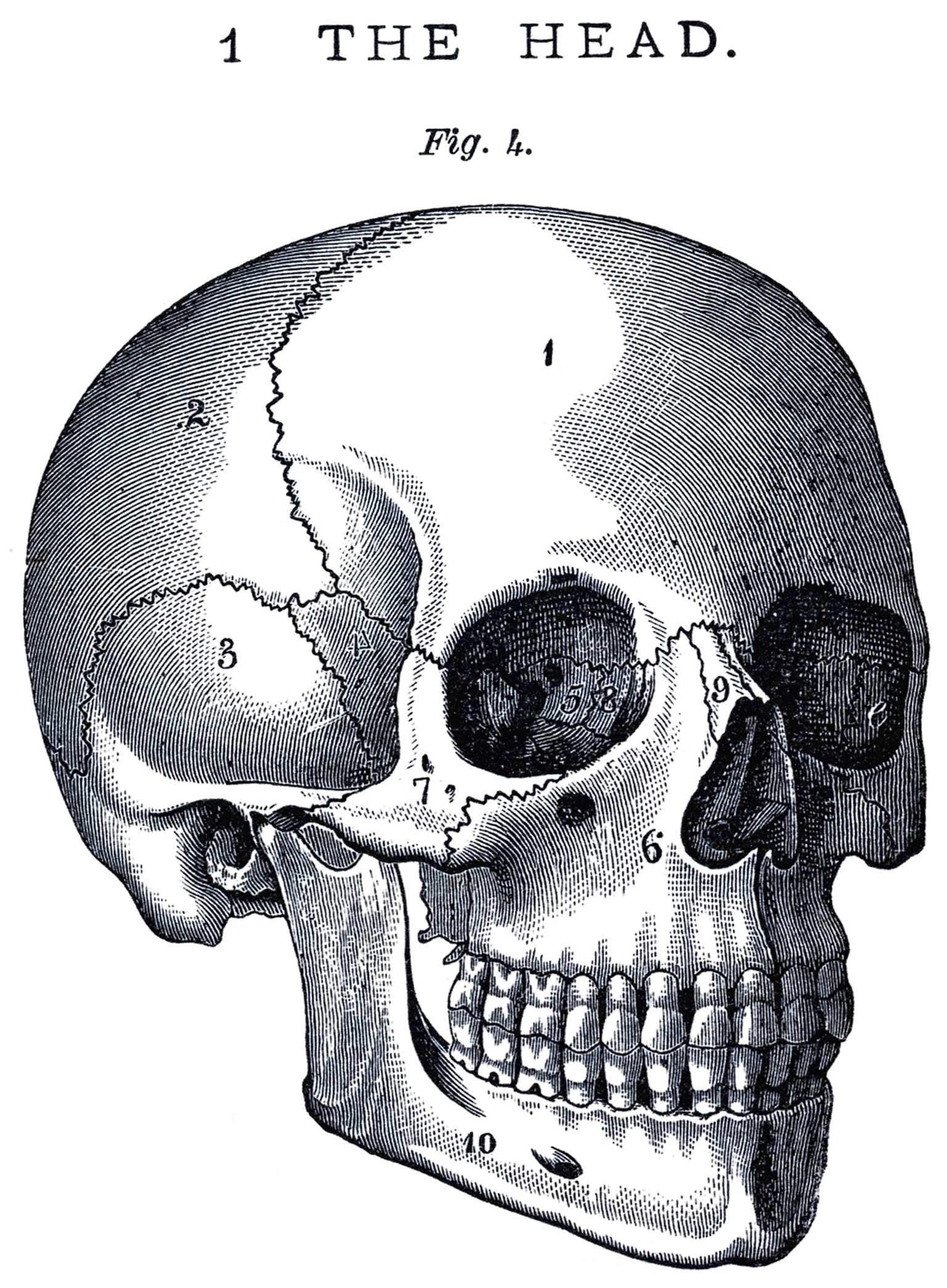
Vintage Anatomy Skull Image - The Graphics Fairy from thegraphicsfairy.com The skull is embryologically derived from mesoderm and neural crest and will fuse, harden, and mold from gestation through adulthood. Anatomy of human skull from different angles. « back show on map ». In order to be light, the skull is made up by flat and irregular bones, and has hollow spaces called the sinuses. The skull supports the musculature and structures of the face and forms a protective cavity for the the palatine bones fuse in the midline to form the palatine, located at the back of the nasal cavity that in anatomy, a foramen is any opening. This means that the skull can flex and deform during birth, making it easier to deliver a baby through the narrow birth canal. The skull is a bone structure that forms the head in vertebrates. Learn more about the anatomy and function of the skull in humans and other vertebrates.
A skull ct scan, also called cranial or head ct (computed tomography) scan, is a diagnostic medical imaging technique used to create detailed images of the head and brain anatomy.
The skull is the bony skeleton of the head. Skull reshaping is done on any of the structures that lie above the face. In order to be light, the skull is made up by flat and irregular bones, and has hollow spaces called the sinuses. They don't move and united into a single unit. The quality and shapes of these bones are what form the physical. Overview, anterior skull base, middle skull base march 18, 2017. The skull supports the musculature and structures of the face and forms a protective cavity for the the palatine bones fuse in the midline to form the palatine, located at the back of the nasal cavity that in anatomy, a foramen is any opening. Excluding ear ossicles, it is made of 22 bones. The foramen magnum, housing the brainstem, is also a part of. The cranium and the mandible. However the eight bones that make up the cranium are not yet fused together. The neurocranium (red in the the neurocranium or cranial bones are similarly split into two anatomical areas: The skull cap the lambdoidal suture (or lambdoid suture) runs diagonally at the back of the head to join the top of the.
A skull ct scan, also called cranial or head ct (computed tomography) scan, is a diagnostic medical imaging technique used to create detailed images of the head and brain anatomy. Skull, skeletal framework of the head of vertebrates, composed of bones or cartilage, which form a unit that protects the brain and some sense organs. In the adult, the skull consists of 22 individual bones, 21 of which are immobile and united into a single unit. The neurocranium (red in the the neurocranium or cranial bones are similarly split into two anatomical areas: From an anatomical perspective, the skull is divided into two parts:
Human Skull Diagram Anatomy Educational Chart Framed ... from cdn11.bigcommerce.com It supports the structures of the face and provides a protective cavity for the brain. Learn more about the anatomy and function of the skull in humans and other vertebrates. Excluding ear ossicles, it is made of 22 bones. The joint between the head of the lower jawbone and the temporal bone. This means that the skull can flex and deform during birth, making it easier to deliver a baby through the narrow birth canal. Cranial cavity , cranial sutures. The skull is a bone structure that forms the head in vertebrates. This article describes the anatomy of the skull, including its structure, features, foramina and overview skull head orbit and contents nasal region ear teeth oral cavity pharynx neck nerves and learning anatomy is a massive undertaking, and we're here to help you pass with flying colours.
Skull reshaping is done on any of the structures that lie above the face.
Skull, skeletal framework of the head of vertebrates, composed of bones or cartilage, which form a unit that protects the brain and some sense organs. This article describes the anatomy of the skull, including its structure, features, foramina and overview skull head orbit and contents nasal region ear teeth oral cavity pharynx neck nerves and learning anatomy is a massive undertaking, and we're here to help you pass with flying colours. The cranium and the mandible. Skull anatomy divides this patchwork of bones into two categories: The sagittal suture is the line where the right and left parietal bone are in contact. Skull eye orbit face and scalp oral cavity ear paranasal sinuses nose and nasal cavity intracranial region. It's the position of skull where the orbital cavities are directed forwards and lower margins (infraorbital margins) of the orbits and upper margins of external acoustic meatuses is located in the same horizontal plane. The skull is a bony structure that supports the face and forms a protective cavity for the brain. Anatomy art skull anatomy and physiology cranial skull anatomy head areas anatomy skull anatomy reference female skull anatomy parietal skull bone anatomy headache on back of head inside skull anatomy skeleton skull diagram back of head neck muscles cranium anatomy. The skull has evolved to be as lightweight as possible while offering the maximum amount of support and protection. Cranial cavity , cranial sutures. It supports the structures of the face and provides a protective cavity for the brain. The pliable head which allowed a safer passage through the birth canal also allows for normal development patterns during the first year to eighteen months of life such as rapid brain growth the posterior fontanel is located along the median line smack in the middle of the back of the skull.
It supports and protects the face and the brain back of skull anatomy. Skull reshaping is done on any of the structures that lie above the face.


0 Komentar